The foetal liver is a critical organ during early development, acting as a blood cell factory before the bone marrow takes over. Here, blood stem cells expand and prepare to support lifelong blood production. At the same time, foetal liver cells mature and develop their metabolic and detoxification functions. Understanding how these different cells work together could provide crucial insights into how we can mimic foetal liver conditions to produce blood outside of the human body, with promising applications in regenerative medicine and transplantation therapies. Read More
Studying the foetal liver presents challenges. Many of its cell types look similar under a microscope, and scientists must stain cells with fluorescent markers in order to distinguish between them. However, there is a lack of specific markers for certain cell populations found in the foetal liver.
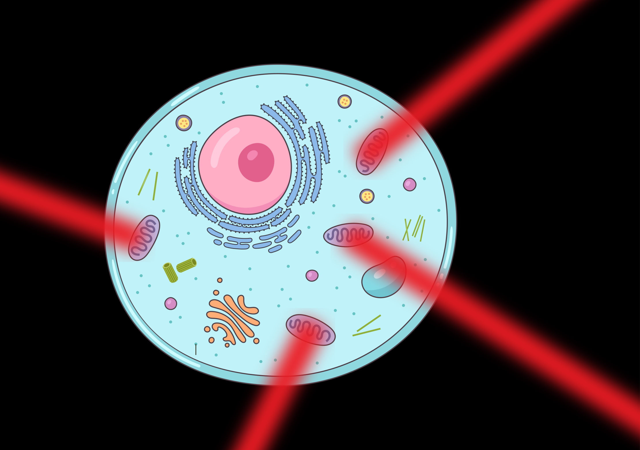
Many foetal liver cells naturally emit light – a phenomenon called autofluorescence. Márcia Mesquita Peixoto, Ana Cumano, and their colleagues at the Institut Pasteur have leveraged autofluorescence as a tool for discovery. Using an innovative technique called spectral flow cytometry, they have identified new cellular characteristics within the foetal liver.
The research team used a high-tech instrument called the Sony ID7000 spectral flow cytometer, which is sensitive enough to detect subtle differences in the way that cells naturally emit light. By analysing light patterns from foetal liver cells, the researchers found distinct autofluorescent signatures that correlate with different cell populations.
One of these signatures identified stellate cells: specialised liver cells that store vitamin A and provide structural support to the liver. The other signature highlighted a group of cells called hepatoblast-like cells, which eventually develop into either liver cells or bile duct cells.
Interestingly, the study revealed that while both hepatoblast-like and bile duct cells emit autofluorescent signals before birth, mature bile duct cells lose this characteristic after birth. This suggests that autofluorescence may serve as a marker for identifying hepatoblast-like cells postnatally – a key breakthrough enabled by spectral flow cytometry.
The team’s discovery allows scientists to study foetal liver development more precisely than ever before. By harnessing autofluorescence using spectral flow cytometry, Cumano, Peixoto Mesquita, and their colleagues have expanded the number of cell types that can be analysed at once, providing a clearer picture of how the foetal liver develops. This new approach could also transform research into other autofluorescent tissues, such as those in the heart, opening up new avenues in biomedical science.