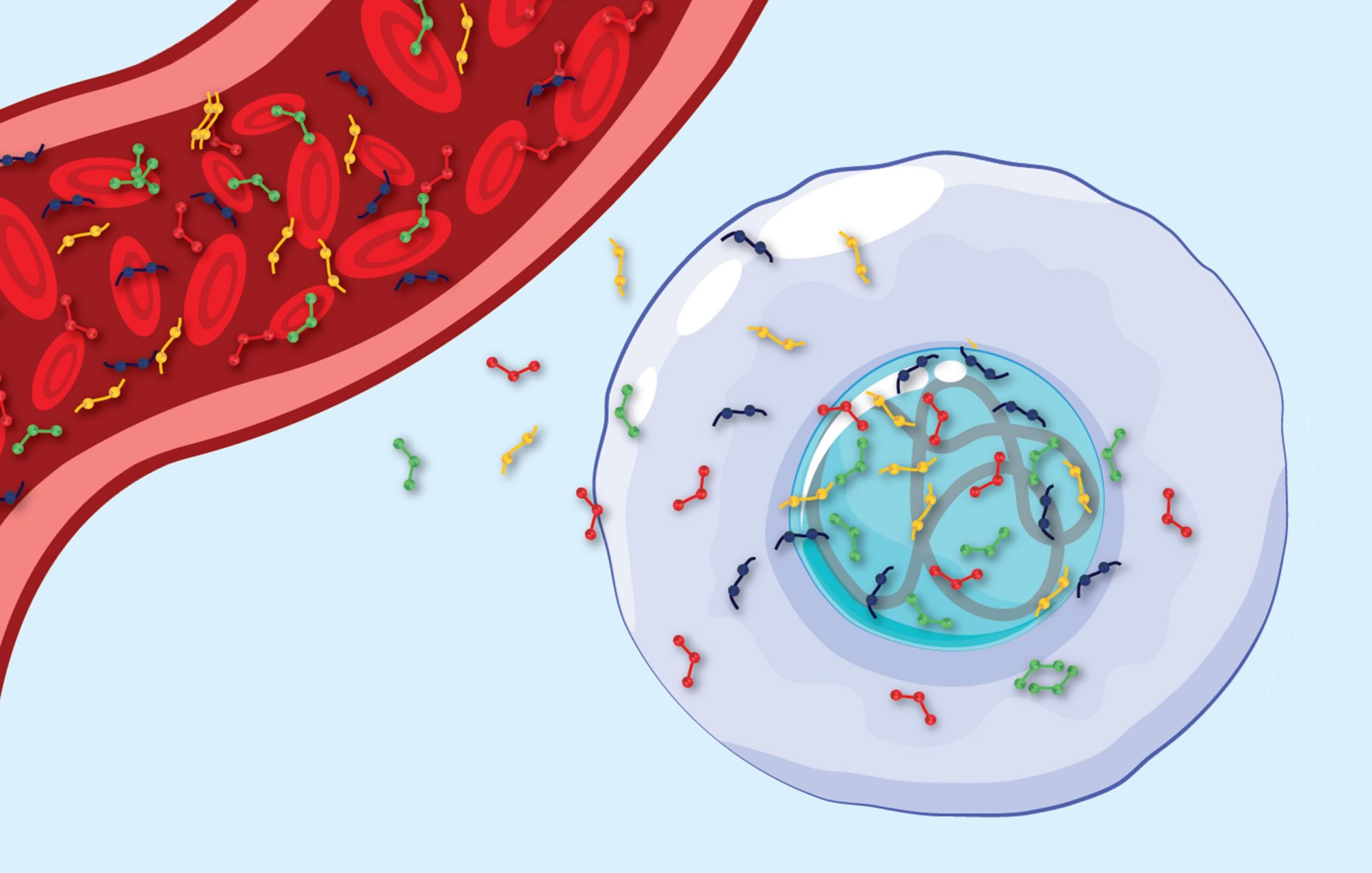
Abdominal aortic aneurysms are a potentially life-threatening condition where the large blood vessel supplying blood to the abdomen and lower body becomes dangerously enlarged. Left untreated, these aneurysms can rupture, often with fatal consequences.
Medical devices called endografts – consisting of a stent and a graft – can be used as internal scaffolds to stabilise the weakened artery walls. However, these devices can lead to problems, which many researchers and clinicians are striving to solve. Read More
Dr Katsuhiko Oda and his team at Iwate Prefectural Central Hospital in Japan have conducted research into one such problem involving the AFX endograft – a medical device that acts like a tube inserted into the weakened artery to prevent it from rupturing.
The AFX endograft is specifically designed for treating abdominal aortic aneurysms by creating a new path for blood flow, bypassing the damaged section of the artery. One standout feature of the AFX endograft is its unibody design, meaning that the stent and graft are integrated into a single, continuous piece rather than being made of connected parts. This design allows it to fit securely in patients with narrow bifurcations – the points where the aorta splits into the arteries that lead to the legs. Narrow bifurcations can make it challenging to use other types of endografts, so the AFX is particularly useful in such cases.
However, Dr Oda’s research revealed that this design can also create long-term challenges. His study, based on two detailed patient cases, uncovers a phenomenon called ‘delayed migration’. Delayed migration occurs when the endograft shifts from its original position years after being implanted, potentially leading to serious complications.
Dr Oda’s team hypothesised that the specific design of the AFX endograft – particularly the flexibility in its lower sections – makes it susceptible to gradual changes under the influence of blood flow and anatomical stress. Specifically, the lower part of the AFX endograft’s main body – the section closest to where it branches into the arteries leading to the legs – is prone to gradually shortening and tilting slightly out of alignment.
This issue is especially common in fusiform-shaped aneurysms, which are balloon-like enlargements of the aorta that expand evenly along the vessel’s length. Over time, these changes can cause the endograft to lose its secure position, leading to complications.
Dr Oda’s team discovered that when the endograft’s main body is slightly angled, the natural flow of blood can exert uneven pressure on certain parts of the device. This pressure can stretch the graft material in certain areas, causing it to bulge and eventually shorten. When the aneurysm itself has a gentle anterior-posterior angulation, this process increases the risk of the endograft shifting position and potentially leaking.
To address this risk, Dr Oda and his colleagues recommend that detailed imaging is carried out before surgery. By using advanced three-dimensional scans to analyse the patient’s anatomy, doctors can better predict whether the AFX endograft is suitable for a specific case.
While the AFX endograft remains a valuable tool for treating certain patients with abdominal aortic aneurysms, Dr Oda’s findings highlight the importance of selecting the right candidates and ensuring long-term follow-up care. By deepening our understanding of the AFX endograft’s strengths and limitations, this study contributes to the ongoing effort to improve outcomes for patients with abdominal aortic aneurysms.